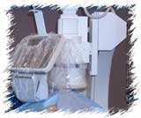
Positioning
of the x-ray camera: The x-ray video camera sits above the patient's
chest, while the x-ray beam is delivered from underneath the table. A
movie camera is attached to the tube to record images on a 35 mm film,
while a cineless lab will forego the movie camera and record the angiogram
on a computer storage drive. Images are also noted "live" on the monitor
and recorded segments can be reviewed and digitally analyzed (catheter
dimension, vessel size, lesion length, ejection fraction, etc.) and manipulated
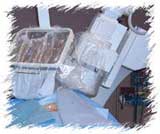
(slow motion, zoom, etc). Positioning
of the x-ray camera: The x-ray video camera sits above the patient's
chest, while the x-ray beam is delivered from underneath the table. A
movie camera is attached to the tube to record images on a 35 mm film,
while a cineless lab will forego the movie camera and record the angiogram
on a computer storage drive. Images are also noted "live" on the monitor
and recorded segments can be reviewed and digitally analyzed (catheter
dimension, vessel size, lesion length, ejection fraction, etc.) and manipulated
(slow motion, zoom, etc).
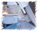
The x-ray tube
is rotated around the patient (side-to-side, and also towards and away
from the head), as shown below. By taking pictures from different angles,
the cardiologist can inspect a stenotic lesion from several points of
view. This increases the accuracy of assessing the clinical importance
and severity of a blockage. It also helps determine the patients candidacy
for angioplasty, stinting, surgery, medical treatment, etc. and in the
selection of the device and its diameter and length (in the case of a
stent).

|