Prior Page Left Coronary Artery RAO View 1 Next Page

 When you have completed review of this screen, please click the "Next" arrow for the second portion of this section.
 
  In the early days of cardiac catheterization, The x-ray tube, image intensifier and camera remained stationary, while the patient lay in a cradle that was rotated from the RAO to the LAO projections. Cranial and caudal angulation were not possible at those times. These "straight" RAO and LAO views were limited because of foreshortening of the left anterior descending and circumflex coronary arteries and the overlap of vessels.
   Today, all cardiac cath labs are equipped with an image intensifier and camera that can be rotated along the RAO/LAO as well as a cranial/caudal (towards and away from the patient's head) projections.  Regardless, the straight RAO may be of occasional value, since it may be the only RAO view that clearly demonstrates the very proximal portion of the LAD or the origin of the obtuse marginal and left postero-lateral branches of the circumflex.

  
In the Right Anterior Oblique or RAO view, the camera is rotated along a vertical axis towards the patient's right, as shown at the bottom of the page. Once again, the size of the heart has been purposely exaggerated for purposes of illustration. Please note that the ventricular septum lies in a plane between the right shoulder and the left nipple. Thus, in the RAO view, the camera "looks" at the outline of the septum. The atrio-ventricular plane is seen on edge, since it sits roughly at right angles to the ventricular septum.

Top of Page

FOR AUDIO: Click the Speaker Icon to "unmute" Audio

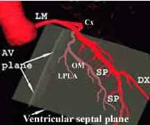
   In the RAO view, the LAD begins close to the spine and then moves away from it and towards the LV apex. It gives off two sets of branches (one or more diagonals and several septal perforators).
 
  The diagonal (Dx) moves diagonally and away from the LAD. The septal perforators (SP) are smaller branches that come off the inferior border of the LAD (at roughly 90 degrees) and travel downward.

  The Cx, in this view,  moves parallel to the spine and give off the obtuse marginal (OM) and left postero-lateral (LPLA) branches that come off at an angle and run roughly parallel to the LAD.
  When you have completed review of this screen, please click the "Next page" blue arrow for the second portion of this section.
Prior Page Left Coronary Artery RAO View 1 Next Page
Main Menu