| Left Coronary Artery RAO View 2 |
The video clip in the middle left panel was obtained in the cardiac cath lab as the left coronary angiogram was recorded in the RAO projection. The movie clip demonstrate both a "video" representation (coronary angios, as they appear on playback in the cath lab) and the "cine" or cineangiographic equivalent as seen on the developed film. The two views correspond to the negative and positive prints of photography film. You can toggle back and forth between the two views by clicking on the respective button. You can also click a button to see a labeled freeze frame of the coronary angiogram. |
| |
 |
|
FOR AUDIO: Click the Speaker Icon to "unmute" Audio 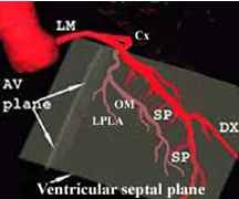 |
 |
  |

| For purposes of reinforcement let
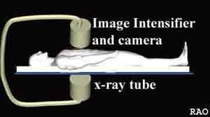
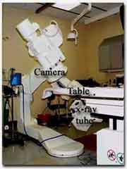
us review the LAD course and branches again. In the RAO view, the LAD begins close to the spine and then moves away from it and towards the LV apex. It gives off two sets of branches (one or more diagonals and several septal perforators). The diagonal (Dx) moves diagonally and away from the LAD. The septal perforators (SP) are smaller branches that come off the inferior border of the LAD (at roughly 90 degrees) and travel downward. The Cx, in this view, moves parallel to the spine and give off the obtuse marginal (OM) and left postero-lateral (LPLA) branches that come off at an angle and run roughly parallel to the LAD. The photograph on the left (above) shows the II and camera assembly in the RAO position. The picture to the right (above) shows the camera's view of the patient's heart. |
| Left Coronary Artery RAO View 2 |