| Left Coronary Artery RAO/Caudal View 2 |
|
FOR AUDIO: Click the Speaker Icon to "unmute" Audio The video clip on the left was obtained in the cardiac cath lab as the left coronary angiogram was recorded in the RAO/caudal projection. The movie clip demonstrate both a "video" representation (coronary angios, as they appear on playback in the cath lab) and the "cine" or cineangiographic equivalent as seen on the developed film. The two views correspond to the negative and positive prints of photography film. You can toggle back and forth between the two views by clicking on the respective button. You can also click a button to see a labeled freeze frame of the coronary angiogram. |

 |
|
 |
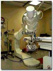
| In the Right Anterior Oblique or RAO-Caudal view, the camera
is rotated along a vertical axis towards the patient's right and also
along the vertical axis away the head and downwards or caudal, as
shown on the right pictures. above. The heart, as "seen" by
the image intensifier/camera assembly is shown on the left (above). Once
again, the size of the heart has been purposely exaggerated
for purposes of illustration. Please remember that the
ventricular septum lies in a plane between the right shoulder and the
left nipple. Thus, in the RAO view, the camera "looks" at the outline
of the septum, but from a point of view that is lower than that of the
RAO view. In other words, the camera looks at the outline of the septum,
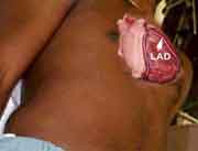
from below. In the RAO-Caudal views, the LAD begins close to the spine
and then moves away from it and towards the left ventricular apex. The diagonal (Dx) moves laterally away from the LAD. The septal perforators (SP) are smaller branches that come off the inferior border of the LAD and travel downwards and medially. The Cx, in the RAO-caudal view, moves away from the LAD and towards the spine before giving off its branches that curve downwards and inferiorly running roughly parallel to the LAD and diagonal coronary arteries. |
| Left Coronary Artery RAO/Caudal View 2 |