| Dominant Right Coronary Artery 2 |
You may click on the buttons to toggle between the LAO and RAO projections, or to have a freeze frame of the angiogram labeled for you. |

|
|
  |
|
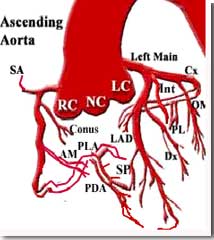
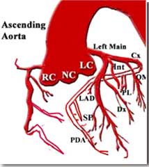
FOR AUDIO: Click the Speaker Icon to "unmute" Audio The diagram on the top left shows a dominant RCA system, as described on the previous screen, while the diagram on the top right shows a non-dominant RCA system. Note that the latter supplies branches to the right ventricle but does not give rise to the PDA, nor does it continue past the AV groove to supply the PLA branchesThe "cine" on the left demonstrates the angiograms of a non-dominant RCA system in the RAO and LAO projections. Click on the buttons at the bottom of the angios to toggle between the two views or to get a labeled freeze frame. |
| Dominant Right Coronary Artery 2 |
