| Right Coronary Artery RAO View 1 |
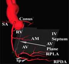
The straight RAO is commonly used in most cardiac cath labs because it clearly demonstrates the mid portions of the RCA, the origin of the acute marginal branch and the length of the posterior descending and postero-lateral branches. It is also a superior view that demonstrates the septal perforators, particularly when it is supplying collaterals to an occluded left anterior descending coronary artery. However, this view suffers from overlap between the posterior descending and postero-lateral branches, a problem that can be solved by using a cranial angulation. |
 |
|
 |
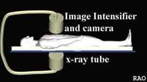
| In the Right Anterior Oblique or RAO view, the camera is
rotated along a vertical axis towards the patient's right, as shown on
the left (above). Please remember that the ventricular septum lies in
a plane between the right shoulder and the left nipple. Thus, in the RAO
view, the camera "looks" at the outline of the septum. The atrio-ventricular
plane is seen on edge, since it sits roughly at right angles to the ventricular
septum, as shown in the animation below. When you have completed review of this screen, please click the "Next page" blue arrow for the second portion of this section. |
| Right Coronary Artery RAO View 1 |
