| Right Coronary Artery RAO/Cranial View 2 |
|
|
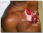
FOR AUDIO: Click the Speaker Icon to "unmute" Audio The video clip on the left was obtained in the cardiac cath lab as the right coronary angiogram was recorded in the RAO/Cranial projection. The movie clip demonstrate both a "video" representation (coronary angios, as they appear on playback in the cath lab) and the "cine" or cineangiographic equivalent as seen on the developed film. The two views correspond to the negative and positive prints of photography film. You can toggle back and forth between the two views by clicking on the respective button. You can also click a button to see a labeled freeze frame of the coronary angiogram. In the RAO/Cranial view, the shaft of the RCA runs parallel to the spine. The right ventricular, acute marginal , posterior descending and postero-lateral branches come off at an angle and travel away from the spine and towards the sternum.
|

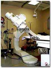  |
| The photograph on the left above shows the x-ray tube, image
intensifier/camera (II) assembly in the RAO/Cranial projection. The tube
is displayed is in a 25 degrees RAO angulation with a 25 degrees cranial
tilt. The photograph on the right shows the camera's view of the patient's
torso and heart. The size of the latter has been exaggerated for purposes
of illustration. |
| Right Coronary Artery RAO/Cranial View 2 |